Serial sectioning transmission electron microscopy (ssTEM)
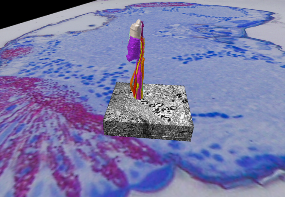
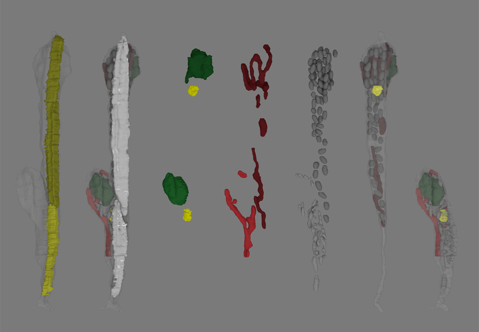
Serial sectioning transmission electron microscopy (ssTEM) is used in order to gain three dimensional information on ultrastructural level by imaging continuous series of ultra-thin sections.
Key publications using ssTEM
Mohr T., Fischer S. (2020) Ultrastructural evidence for the origin of the sub-retinal pigment shield in the compound eye of Drosophila melanogaster. J. Morph. https://doi.org/10.1002/jmor.21143
Mohr T., Meinertzhagen I.A., Fischer S. (2019) Novel type of sub‐retinal pigment shield in the miniaturized compound eye of Trichogramma evanescens. J Comp Neurol https://doi.org/10.1002/cne.24745
Fischer S., Lu Z., Meinertzhagen I.A. (2019) Three-dimensional ultrastructural organization of the ommatidium of the minute parasitoid wasp Trichogramma evanescens. Arthropod Structure and Development (48:35-48). https://doi.org/10.1016/j.asd.2018.12.003
Fischer S., Lu Z., Meinertzhagen I.A. (2017) From two to three dimensions: The importance of the third dimension for evaluating the limits to neuronal miniaturization in insects. J Comp Neurol. (526(4):653-662). https://doi.org/10.1002/cne.24358